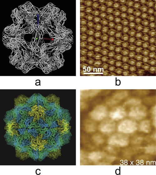
Fig. 8.
(a) Polypeptide backbone structure, determined by X-ray crystallography, of the T = 1 particle that forms when the T = 3 virion of brome mosaic virus is treated with high salt and neutral pH. It is seen looking along a 2-fold axis. (b) Surface of a crystal of the BMV T = 1 particles. (c) Polypeptide backbone structure of the T = 3 icosahedral turnip yellow mosaic virus, also determined by X-ray diffraction analysis. (d) A single virion of TYMV, imaged by AFM, which was incorporated into the surface of a crystal of the virus. The pentameric and hexameric capsomeres are evident in the AFM image.

