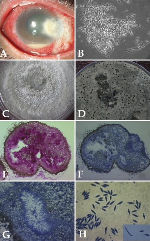
Fig. 1.
(A) Slit lamp photograph showing infected cornea involving regions of sclera. (B) Ten percent KOH mount of the scraping material showing fungal hyphae (magnification, ×400). (C) Growth of case isolate P. phoenicicola on PDA showing flat, spreading colony with gray-white sparse aerial mycelium. (D) Growth of case isolate P. phoenicicola on rice flake agar showing pycnidia (black dots) after 4 weeks at 25°C. (E to G) Histological sections of thick-walled pycnidium. (E) Staining with PAS (magnification, ×100). (F) Staining with lactophenol cotton blue (magnification, ×100). (G) Alpha and beta conidia lining the cavity (magnification, ×400). (H) Alpha and beta conidia (magnification, ×1,000).

