Fig.2.
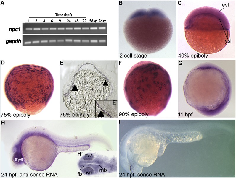
Expression of zebrafish npc1 during embryogenesis. (A) RT-PCR analysis of zebrafish npc1 from embryos aged from 1 hpf to 7 dpf (RT samples showed no signal, not shown). (B–H) Lateral (B–I) or dorsal (H′) views of embryos stained with a RNA probe for npc1, (B) in blastodiscs at the 2 cell stage, (C–F) during epiboly stages in the blastomeres extending from the animal to vegetal poles and in the YSL. Expression in the YSL tissue was confirmed in embryo sections (arrowheads in E) and at higher magnifications of 75% epiboly sectioned embryo (E′). (G) During early somitogenesis npc1 was ubiquitously expressed at low levels. (H, H′) By 24 hpf, npc1 became mostly localized to the anterior tissues, with strong staining in the neural tissues, as visible in ventral views of stained heads (H′). (I) Embryos stained at 24 hpf with a npc1 sense RNA probe had no detectable signal. fb, forebrain; mb, midbrain.

