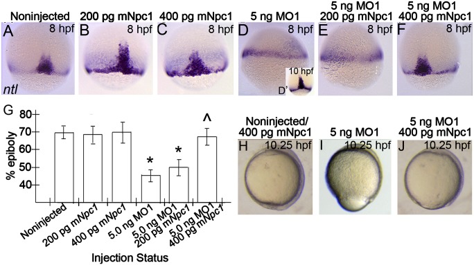
Fig.4.
Epiboly-delay phenotypes in npc1 morphants are rescued by co-injection with mouse Npc1 mRNA. (A–F) Lateral views, animal pole facing upward of fixed noninjected or injected embryos stained with ntl riboprobe at 8 hpf, or MO1-injected embryos at 10 hpf (D′). (A–D) Embryos injected with mNpc1 mRNA alone were indistinguishable from noninjected controls (A–C), while the blastoderm margin of time-matched embryos injected with MO1 had progressed at a lower rate by 8 hpf (D), but continued to progress and looked similar to 8 hpf noninjected controls by 10 hpf (D′). (E, F) Co-injection of 400 pg mNpc1, but not 200 pg mNpc1, rescued MO1-associated defects. (G) The effect of expressing mNpc1 RNA in wild-type and MO1-injected embryos. Graph represents the % of epiboly from averages obtained on analyzing the epiboly progression in each group from a single clutch of siblings. Error bars show standard deviation. Statistics derived from posthoc analysis after ANOVA. *P < 0.001 compared with control embryos; ^P < 0.001 compared with MO1 injection alone. (H–J) Lateral views, anterior structures facing upward of live controls or mRNA injected embryos (H), MO1-injected embryos (I), or embryos co-injected with RNA and MO1 (J) at 10.25 hpf.

