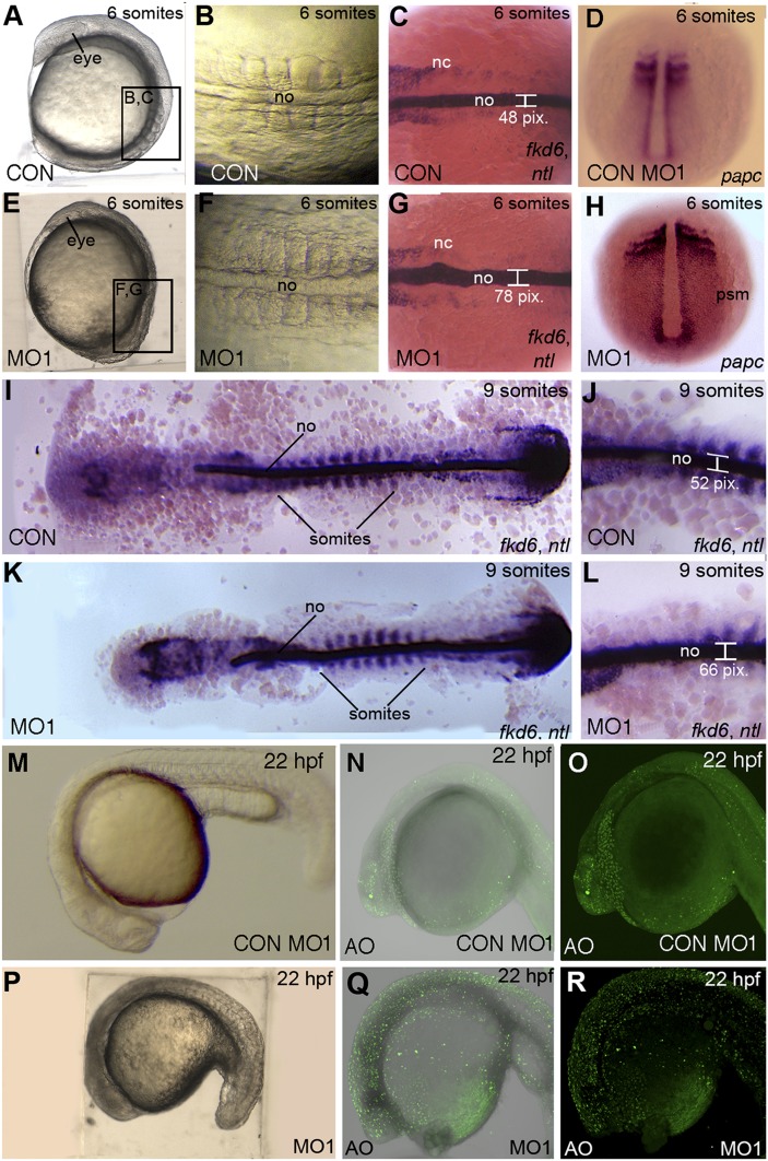
Fig.9.
The npc1 morphants display body axis defects and cell death. Lateral views of six-somite stage (A, E) and 22 hpf (M–R), or dorsal views of six-somite stage (B, C, D, F, G, H) and nine-somite stage (I–L) control (A–D, I, J, M) or npc1 morphants (E–H, K, L, N, O). (A, B, E, F) Nomarski optics reveal that npc1 morphants have a shorter and wider body axis than stage-matched controls at the six-somite stage. (C, D, G–L) Staining the midline mesoderm with an RNA probe for ntl revealed a widening of npc1-morphant midline (width 70 ± 5.1 pixels; n = 14) compared with controls (width: 56 ± 7.1 pixels; n = 11; P < 0.01) while papc staining revealed widening of the lateral mesodermal structures. (I, K) Deyolked and flat-mounted npc1 morphants are shorter in the anterior-posterior direction at the nine-somite stage. ntl staining marks nine somites in each embryo for proper stage matching. (J, L) ntl-stained npc1 morphants display a wider notochord at nine somites (64 ± 5.7 pixels; n = 12) compared with controls (52.6 ± 5.2 pixels; n = 10; P < 0.01. (M, P) By 22 hpf, MO1-injected embryos had darkened populations of cells in anterior regions, suggestive of cell death. (N, O, Q, R) Brightfield and fluorescent images of acridine orange (AO)-stained embryos. The npc1 morphants displayed increased levels of cell death, compared with Con MO-injected siblings. nc, neural crest; no, notochord; psm, presomitic mesoderm.

