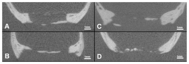
Figure 8.
Cross sections thru the center of the defect using 3D microCT evaluation. All samples are in vivo scans of solid 100 μm scaffolds following 90 days healing. A: Healing predominantly along periosteal side. B: Healing predominantly along dural side. C: Healing along both periosteal and dural sides. D: Healing located centrally within defect.

