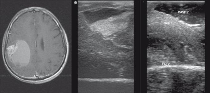
Figure 4.

Right parietal recurrent oligodendroglioma. MRI (left panel) showing a predominantly cystic lesion with a peripheral solid area. Preresection IOUS (centre panel) depicting the lesion and postresection (right panel) IOUS showing the resection cavity.
