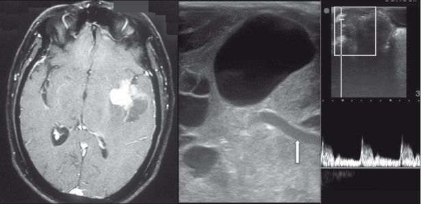
Figure 6.

Sylvian fissure mass. Contrast axial MRI showing a heterogeneous lesion in the sylvian fissure (left panel). IOUS image showing the underlying middle cerebral artery (arrow, center). Post-resection Doppler showing patent vessels (right panel).
