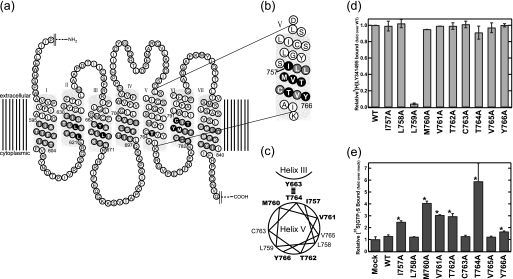
FIGURE 1.
Constitutively active mutations in helix V of mGluR8. a and b, shown is a two-dimensional diagram of mGluR8. The residues that were replaced by alanine (or serine) in the previous (13) (a) and present (b) alanine-scanning mutagenesis are indicated by either black or gray shading. Black shading indicates the residues whose substitution resulted in an increase of constitutive activity. c, shown is helical wheel projection of constitutively active mutation sites in helix V. Residues whose substitution resulted in an increase of basal activity are indicated by bold letters. d, ligand binding potency of alanine mutants in helix V is shown. [3H]LY341495 binding to wild-type and mutant-expressing HEK293 cell membranes was measured. Results are normalized to total specific binding of wild-type. e, constitutive activity of alanine mutants in helix V are shown. The asterisk indicates a significant increase of constitutive activity in the mutants (p < 0.05; Student's t test, two-tailed). All data were normalized to the activity of the mock-transfected membranes and are expressed as the mean ± S.D. of more than two independent experiments done in duplicate.

