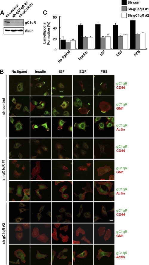
FIGURE 3.
Knockdown of gC1qR prevents growth factor-induced lamellipodia formation. A, expression of gC1qR was determined by immunoblot analysis of whole cell lysates obtained from a A549 clone expressing a scrambled control shRNA (sh-con), and two A549 clones stably expressing shRNA for gC1qR (sh-gC1qR #1 and sh-gC1qR #2). Actin was used as a loading control. B and C, after 18 h of serum starvation, sh-con and sh-gC1qR cells were stimulated with serum or growth factors as indicated in Fig. 1B. After permeabilization, the cellular colocalization of gC1qR with CD44, actin, and GM1 was determined by immunofluorescence using anti-gC1qR, anti-CD44, rhodamine-conjugated phalloidin, and rhodamine-conjugated CTB, respectively (B). Scale bar, 20 μm. The supplemental Fig. S2 shows the fluorescence imaging of each molecule. The ratio of the number of cells with GM1-containing lamellipodia to the total number of cells is given (C).

