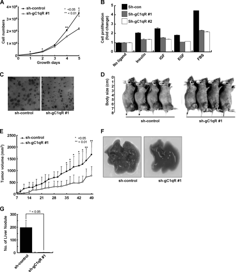
FIGURE 7.
Knockdown of gC1qR abolished tumorigenesis and metastasis of A549 cells in nude mice. A, proliferation of sh-con and sh-gC1qR #1 A549 cells. Both cell lines were seeded at 5 × 10⁴ cells per 60-mm dish. The total cell number was counted on the indicated days. B, growth factor-dependent proliferation of sh-con and sh-gC1qR A549 cells. These cell lines were seeded at 5 × 10³ cells per 96-well plate and cultured for 48 h. The cells were serum-starved for 18 h and then treated with serum or growth factors. After 72 h, the MTT assay was performed as described under “Experimental Procedures.” Cell proliferation was expressed as a fold change relative to nontreated cells. C, anchorage-independent growth of sh-con and sh-gC1qR #1 A549 cells on soft agar. Photomicrographs of the colonies were obtained 3 weeks after plating (×40). D and E, photographs (D) and measurement of tumor volume (E). sh-con or sh-gC1qR #1 A549 cells (4 × 10⁶ cells) were subcutaneously injected into the right hind legs of BALB/c athymic mice (n = 4). Photographs were taken on day 49, and the tumor volume was measured every other day until day 49. (*, p < 0.05, n = 4 per group.) Filled circle, sh-con; open circle, sh-gC1qR #1. F and G, macroscopic observation (F) and quantification (G) of liver metastasis. sh-con or sh-gC1qR A549 cells (5 × 10⁵ cells) were injected into the tail veins of BALB/c athymic mice (n = 5), and liver nodules were counted 28 days post-injection. (*, p < 0.05, n = 5 per group.)

