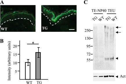
FIGURE 6.
Expression of Flg2 in the epidermis of wild type and calpastatin transgenic mice. A, representative sections of paraffin-embedded skin of wild type (WT) and calpastatin-overexpressing transgenic (TG) mice were analyzed by indirect immunofluorescence with anti-Flg2 antibodies. Flg2 was located in the granular cells and the lower corneocytes. The dotted lines indicate the dermo-epidermal junction. Scale bars, 10 μm. B, the intensity of detection was quantified (n = 4) and shown to be higher in the TG mice. *, p < 0.05. C, epidermal proteins from WT and transgenic mice were sequentially extracted in isotonic buffers containing either Nonidet P-40 (TE-NP40) or 8 m urea (TEU). Equal volumes of each extracts were separated on 7.5% acrylamide gels, and proteins were immunoblotted with the anti-Flg2 antibodies (top) and a monoclonal antibody directed against actin (Act; bottom). A picture representative of at least three different experiments is shown. In the upper panel, the arrows indicate the full-length Flg2 and a processed form of 200 kDa, and the arrowhead indicates a fragment of 55 kDa. Molecular masses of standards are indicated in kDa to the left.

