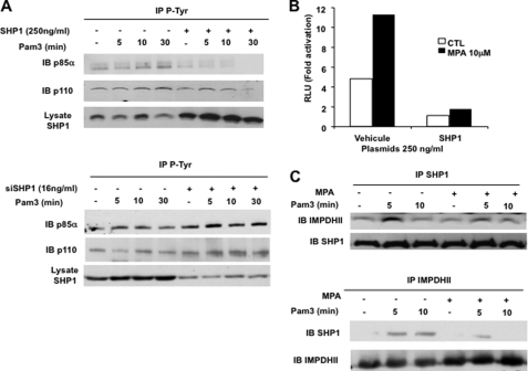
FIGURE 8.
IMPDHII interacts with SHP1, which negatively regulates TLR2-mediated phosphorylation of PI3K. A, HEK293-T2 cells were transfected with SHP1 expression vector (250 ng/ml) (upper panel) or siRNA targeting SHP1 (16 ng/ml) (lower panel) and stimulated with Pam3 (1 μg/ml). Cells were then stimulated with Pam3 and immunoprecipitated with anti-phosphotyrosine antibodies (4G10 clone) (IP). The P85α and p110 subunits of PI3K were visualized by Western blot (IB). B, HEK293-T2 cells were co-transfected with SHP1 expression vector (250 ng/ml), 5× κB-luciferase promoter gene plasmid (40 ng/ml), and β-galactosidase expression vector (40 ng/ml). Cells were then incubated or not with MPA for 2 h and stimulated with Pam3 for 6 h; NF-κB-driven luciferase activity was measured and rationalized upon β-galactosidase expression. Results are expressed as arbitrary units (RLU, relative luciferase units) corresponding to “-fold activation.” C, THP1 cells were exposed to MPA (10 μm) or DMSO for 2 h. Cells were then stimulated with Pam3 (1 μg/ml) and immunoprecipitated with 3 μg of anti-SHP1 or anti-IMPDHII antibodies. IMPDHII and SHP1 were detected by Western blot. Results are representative of three independent experiments.

