FIGURE 5.
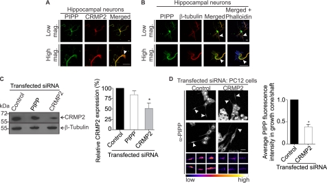
CRMP2 and PIPP co-localize in the growth cone and axon shaft and PIPP growth cone localization is CRMP2 dependent. A, hippocampal neurons were stained with PIPP (PIPP-AB1) and CRMP2 antibodies and imaged by confocal microscopy. Arrowhead shows colocalization of PIPP and CRMP2 in the growth cone. Higher power images are shown below. Bar = 20 μm. B, hippocampal neurons were costained with PIPP (PIPP-AB1) and β-tubulin antibodies and with phalloidin (blue) staining. Colocalization of β-tubulin and PIPP is indicated by arrowheads. Higher magnification images are shown below. Bar = 20 μm. C, PC12 cells expressing control, PIPP, or CRMP2 siRNA were immunoblotted with CRMP2 antibodies, then re-probed with β-tubulin antibodies. Relative CRMP2 expression was determined by densitometry and expressed as a % of the scrambled, designated 100%. Bars = mean ± S.E. (n = 5, *, p < 0.05). D, cells transfected with control or CRMP2 siRNA were differentiated for 3 days and costained with PIPP antibodies (PIPP-AB4) and imaged by confocal microscopy at the same laser attenuation. Arrowheads indicate growth cones of Cy3 siRNA-transfected cells. Bar = 20 μm. The smaller panels below show representative examples of additional growth cones imaged at the same laser attenuation and at higher magnification for each indicated transfected siRNA. Growth cones are presented in graded fluorescence. Bars = mean fluorescence intensity of PIPP in the growth cone relative to the neurite shaft ± S.E., where the control is designated 1 (≥10 cells, n = 3, *, p < 0.05).

