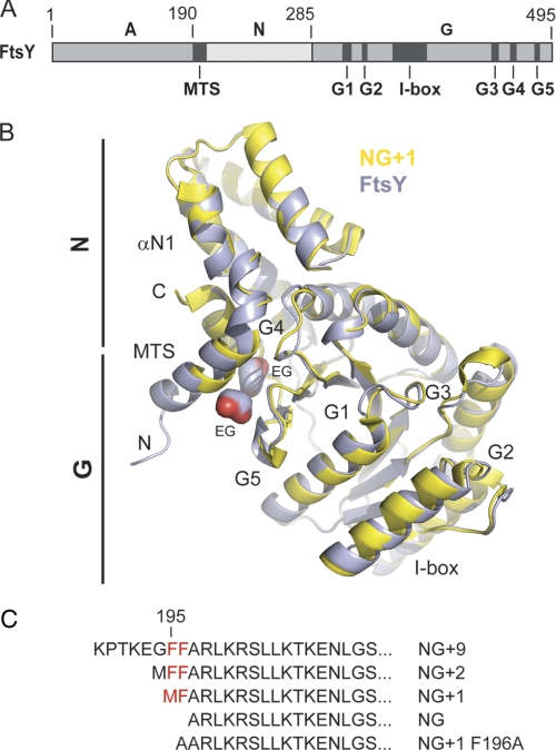
FIGURE 1.
Crystal structure of FtsY allows definition of complete MTS. A, the domain architecture of the E. coli SRP receptor FtsY. The location of the MTS, the I-box, and the conserved G elements (G1–G5) are indicated. B, superposition of FtsY (blue) and NG+1 (yellow; Protein Data Bank code 2QY9) crystal structures. FtsY and NG+1 are shown in ribbon representation. The MTS present at the N terminus of αN1 is extended in the FtsY structure by two turns (residues 188–195). N and C termini of FtsY are indicated by “N” and “C,” respectively. Two ethylene glycol molecules associated to the hydrophobic face of the MTS are indicated by “EG.” C, N-terminal sequences from various FtsY variants used in this study. The conserved double phenylalanine motif is colored in red.

