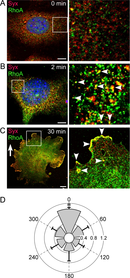
FIGURE 2.
Syx and RhoA colocalized in response to VEGF. A, there was little colocalization of Syx and RhoA in quiescent ECs. B, Syx and RhoA colocalized on endocytic vesicles within 2 min of stimulation by 20 ng/ml VEGF-A164. C, Syx and RhoA polarized in the direction of the VEGF-A164 gradient (0–20 ng/m) and colocalized on endocytic vesicles. D, quantification of the extent and angular distribution of Syx and RhoA colocalization in ECs subjected to a VEGF-A164 gradient (0–20 ng/m). The gradient direction is 0 degrees (mean ± S.D., n = 10; p < 0.0001). Bars, 10 μm.

