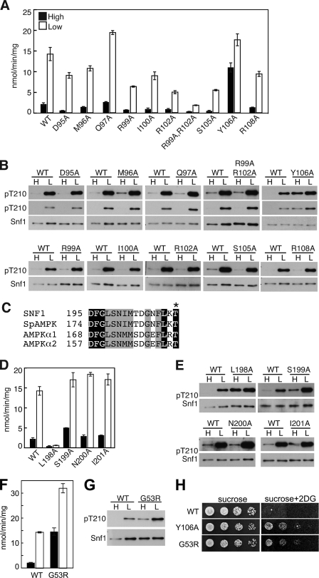
FIGURE 2.
Residues in the Snf1 kinase domain affect SNF1 activity and phosphorylation. A, B, and D–G, WT and mutant Snf1 proteins were expressed in snf1Δ cells. Assays of catalytic activity and immunoblot analysis were as in Fig. 1. Immunoblot analysis of three transformants expressing each mutant protein gave similar results. C, sequence alignment of the DFG motif and following residues for yeast and human members of the SNF1/AMPK family. Conserved residues are highlighted. Asterisk, phosphorylated Thr. H, cells were grown overnight in selective SC plus 2% glucose and spotted with serial 5-fold dilutions on selective SC solid medium containing 2% sucrose or 2% sucrose plus 200 μg/ml 2-deoxy-D-glucose (2DG). Plates were incubated at 30 °C for 2 or 3 days, respectively, and photographed. Rows were from the same plates.

