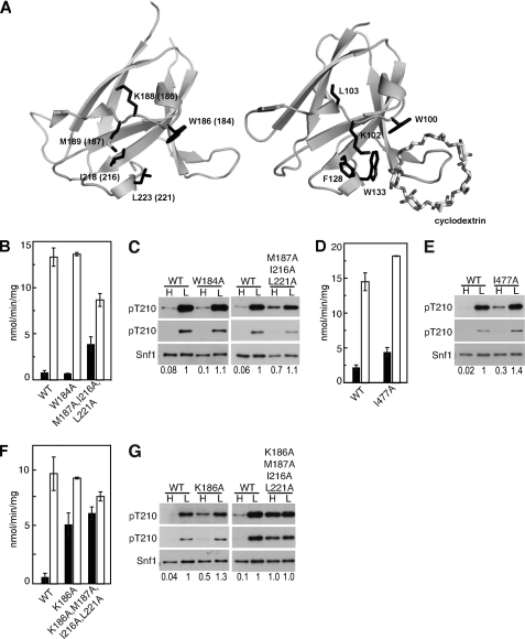
FIGURE 4.
Residues in the Gal83 GBD are required to maintain SNF1 in the dephosphorylated state in high glucose. A, schematic representation of the structures of Sip2 GBD (PDB accession code 2QLV) (30) with corresponding Gal83 residue numbers in parentheses, and mammalian GBD bound to β-cyclodextrin (PDB accession code 1Z0M) (54). For residues mentioned in the text, the corresponding side chains are shown for each structure. The structure figures were produced with PyMOL (DeLano, W. L. (2002) The PyMOL Molecular Graphics System, DeLano Scientific LLC, San Carlos, CA). B–G, assays of catalytic activity and immunoblot analysis as in Fig. 1. B, C, F, and G, Gal83 proteins were expressed in gal83Δ sip1Δ sip2Δ cells. D and E, Snf1 proteins were expressed in snf1Δ cells. Immunoblot analysis of three transformants gave similar results. Values indicate relative intensity of the bands corresponding to phosphorylated Snf1-Thr210 and total Snf1 protein on the longer exposures.

