FIGURE 3.
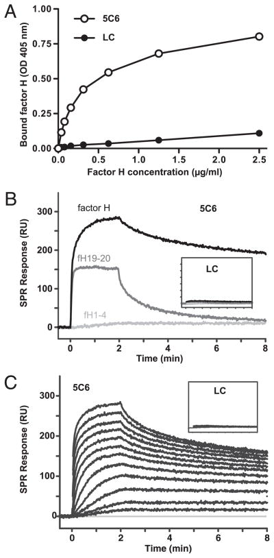
Binding of factor H to surfaces coated with synthetic 5C6 peptide as assessed by ELISA (A) and SPR (B, C). A, Polystyrene wells were coated with streptavidin, saturated with BSA, and incubated with biotinylated 5C6 peptide or negative control peptide; after incubation with serially diluted factor H, bound factor H was detected by a polyclonal Ab. Data shown are means ± SD (within the symbols) from a representative of three experiments. B and C, Biotinylated 5C6 peptide or control (LC, insets) was captured on streptavidin-coated SPR sensor chips, and factor H fragments at a fixed concentration (100 nM; B) or a dilution series of factor H (0.24–500 nM; C) was injected for 2 min with a dissociation phase of 6 min. The data are representative of two independent experiments on different SPR instruments showing comparable results. Additional evaluation of SPR data can be found in Supplemental Fig. 1. LC, linear compstatin; RU, resonance units.

