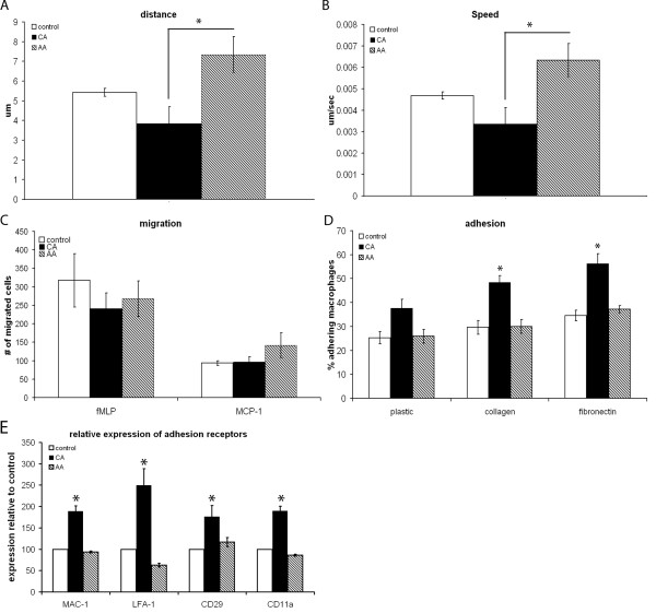
Figure 3.
Motility and migratory capacity. Motility of in vitro stimulated bone marrow derived macrophages was determined by timelapse video microscopy. Speed and distance covered were determined using TrackIt software. The migration towards fMLP and MCP-1 was used as a measure for migratory capacity of in vitro stimulated bone marrow derived macrophages. Adhesion was determined by plating fluorescently labeled macrophages on (un)coated 96 wells plates, washing the plates after 2 h and measuring fluorescence. Data are the mean of 3 separate experiments (n = 3) ± SEM. *= p < 0.05. A) During the recorded time the average distance AA macrophages covered was significantly enhanced twofold compared to CA macrophages. The distance was recorded in μm. B) AA macrophages had a twofold higher speed compared to CA macrophages. Speed was recorded in μm/sec. C) No difference in migration was seen between the different macrophage phenotypes when comparing migration towards either fMLP or MCP-1. D) CA macrophages displayed a trend towards higher adherence on plastic compared to control and AA macrophages. On fibronectin and collagen adherence of CA macrophages was twofold higher compared to both control and AA macrophages. E) The expression of adhesion receptors was higher in CA macrophages compared to both AA and control macrophages.

