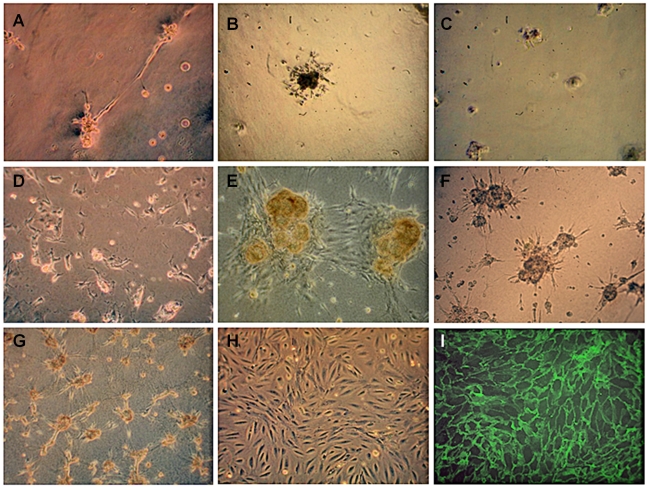
Figure 4. Appearance of GEnC cultured for 1 week at 33°C on various matrices to identify a suitable matrix to support growth of GEnC in a uniform layer by light (A–H) or immunofluorescence (I) microscopy.
(A) GEnC formed capillary tube-like structures on reduced growth factor (RGF) Matrigel, (B) clumps on RGF Matrigel coated with fibronectin or (C) gelatin, (D) rounded up on peptide hydrogel, (E) formed clumps on collagen type I and on (F) collagen type I coated with 20 µg/ml fibronectin or (G) gelatin. (H) GEnC formed a confluent monolayer on tissue culture plastic and on (I) Cellagen (visualised using IF staining for VE-cadherin, as Cellagen is opaque). All cells were seeded at the same density. Original magnification ×10.

