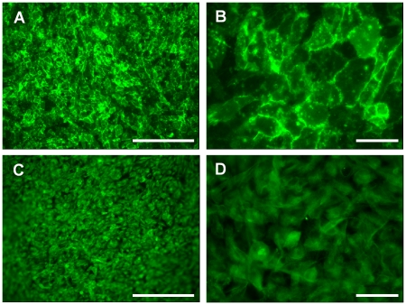
Figure 5. Immunofluorescence staining of (A and B) GEnC and (C and D) podocytes demonstrating monolayer formation when cells are cultured on collagen/PCL electrospun membranes for 1 week at 33°C.
GEnC are stained for PECAM-1 and podocytes for podocin. Scale bars bar 250 µM (A and C) and 50 µm (B and D).

