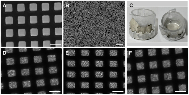
Figure 7. Illustrative images of the micro-PEF nickel mesh and the mesh coated with electrospun nanofibres to form a bioartificial composite membrane.
(A) Light microscopy of micro-PEF nickel mesh (scale bar 25 µm). The black lines are the bars of the mesh, white squares are the open area. (B) Scanning EM of the micro-PEF nickel mesh coated with electrospun collagen/PCL nanofibres, scale bar 10 µm. (C) Collagen I/PCL/nickel mesh bioartificial composite membrane secured in 10 mm diameter Cell Crowns. (D,E and F) Light microscopy of the collagen/PCL/mesh composite after 1, 3 and 5 days incubation in tissue culture media at 33°C (scale bar 25 µm).

