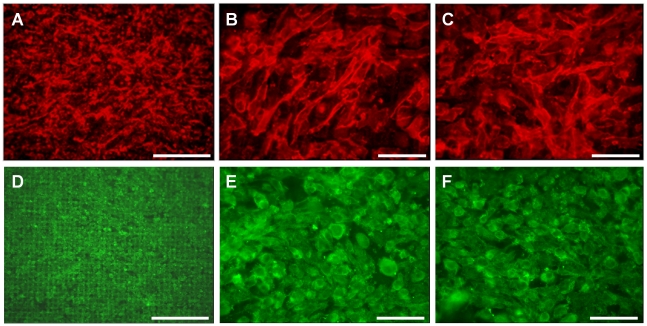
Figure 9. Immunofluorescence demonstrating appearance of GEnC (A–C) and podocyte (D–F) cell layers after one week at 33°C and one week at 37°C on the collagen/PCL/mesh bioartificial composite membrane.
PECAM-1 on GEnC labelled red and podocin on podocytes green. (A and B) GEnC cultured in a single layer or (C) with podocytes co-cultured on the opposite side of the membrane. (D and E) podocytes cultured in a single layer or (C) with GEnC co-cultured on the opposite side of the membrane. Scale bars 250 µm (A and D) and 50 µm (B, C, E and F).

