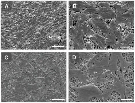
Figure 10. Scanning EM demonstrating morphology of GEnC (A and B) and podocytes (C and D) co-cultured on opposite sides of the collagen/PCL/mesh bioartificial composite membrane.
Scale bars 100 µm (A and C) and 20 µm (B and D). GEnC form a uniform monolayer, comparable with those grown in monoculture or co-culture with podocytes on porous supports in tissue culture inserts (Fig. 2), whilst podocytes are less densely packed.

