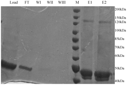
Figure 3. The on-column glutaraldehyde crosslinking of the mycobacterial CoaE.
The 8% SDS-PAGE gel picture shows the CoaE protein loaded on the column (Load); the flowthrough from the column during loading (FT), the eluate during the wash steps (WI, WII and WIII); the molecular weight marker (M) and the elution aliquots (E1 and E2).

