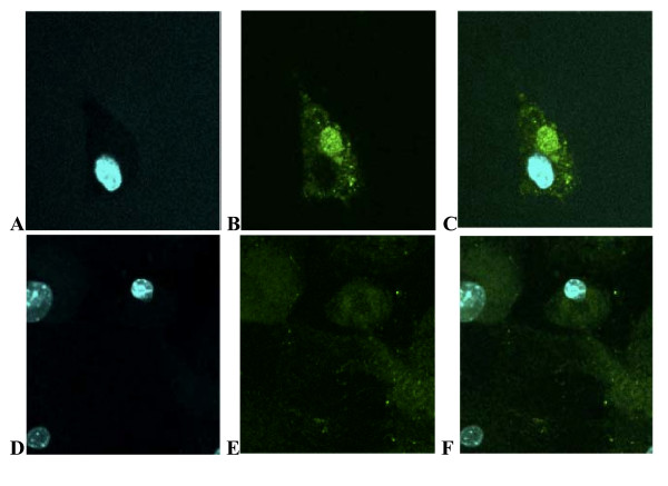
Figure 1.
Catalase is targeted to mitochondria of tumor cells from mCAT positive PyMT mice. Mammary tumor cells stained with human primary anti-catalase and Alexa 488 secondary antibodies and counter-stained with Hoechst 33342 staining for the nucleus. A-C showing nuclear stain, mitochondrial catalase stain and overlap in PyMT cells expressing mCAT and D-F in PyMT cells negative for mCAT, respectively.

