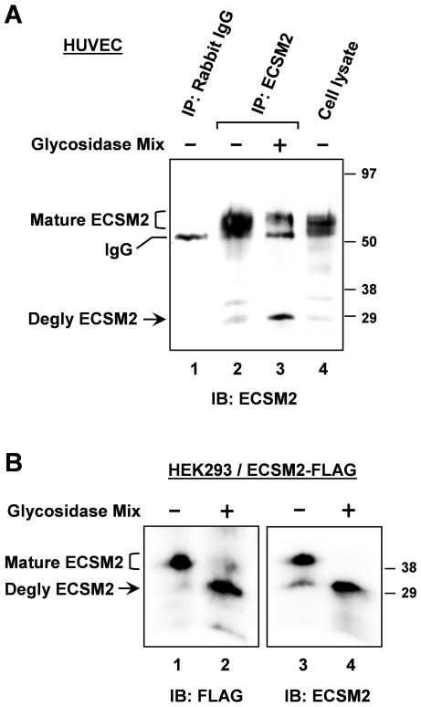
Figure 2. Characterization of ECSM2 proteins by enzymatic deglycosylation.
(A) Glycosylation of endogenous ECSM2. HUVEC lysates were immunoprecipitated with anti-ECSM2 RabMAb (lanes 2 and 3) or rabbit IgG as a control (lane 1). Samples were treated with (+) or without (-) glycosidase mix, as detailed in Methods, resolved by SDS-PAGE, and immunoblotted with anti-ECSM2 RabMAb. Positions of glycosylated (mature) ECSM2, deglycosylated ECSM2, and IgG are indicated. (B) Glycosylation of ECSM2-FLAG. HEK293 cells stably expressing mouse ECSM2-FLAG were lysed and cell lysates were directly subjected to enzymatic deglycosylation reactions as described in Methods. Samples were analyzed by immunoblotting with anti-FLAG M2 mAb (lanes 1 and 2) and anti-ECSM2 RabMAb (lanes 3 and 4), respectively. Positions of glycosylated (mature) ECSM2-FLAG, and deglycosylated ECSM2-FLAG are indicated.

