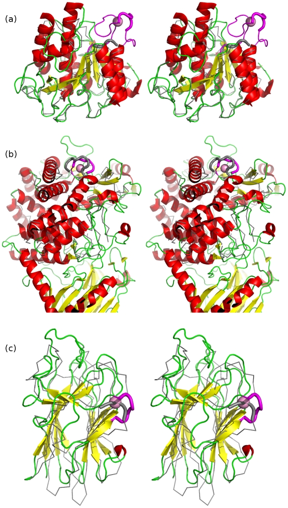
Figure 2. Stereo structure superpositions of novel Dx[DN]xDG calcium-binding motifs with nearest non-calcium binding structural neighbours.
Panel a) shows T. kodakaraensis subtilisin (PDB code 2z2x), b) E. coli YgjK (PDB code 3c68) and c) the Porphyromonas adhesion domain (PDB code 3km5). In each case the Dx[DN]xDG motif is shown as a thick magenta cartoon with bound calcium in pink and the remainder of the calcium binding protein coloured by secondary structure. In a) the Dx[DN]xDG motif is positioned in a larger insertion binding four calcium ions which is also shown in magenta. Structural neighbours (Bacillus lentus subtilisin (PDB code 1c9m) in a), a predicted hydrolase from Thermus thermophilus (PDB code 2z07) in b), and an adhesion domain from human Tyr phosphatase mu (PDB code 2v5y) in c) are in grey with the portion aligning to the calcium binding region shown as thick cartoon. Note that the fourth novel context (2zxq) has no non-calcium binding structural neighbour in the present PDB.

