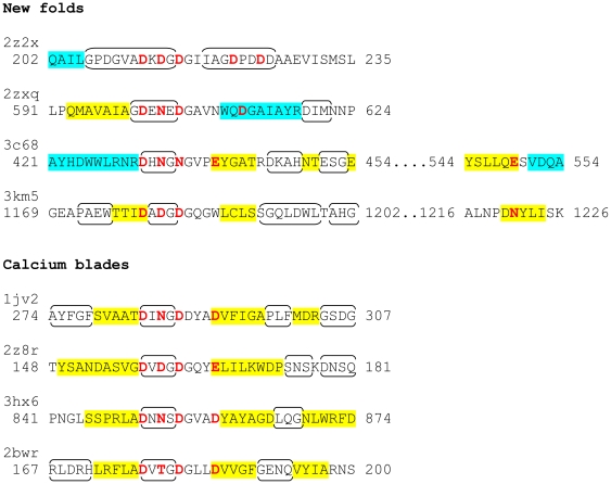
Figure 3. Secondary structure context of the Dx[DN]xDG motifs, highlighting additional metal-binding residues ( Table 1 ).
Residues binding to metal using side chains are in red (direct interaction with calcium) or purple (through-water interaction). Secondary structure as defined by STRIDE [78] is indicated as follows: α-helices, blue shading; β-strands, yellow shading; turns, brackets. A version including previously reported families is included as Figure S1.

