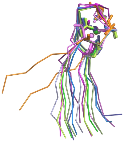
Figure 4. Comparison of calcium blades and their flanking β-strands.
Backbone is shown as ribbon, side chains that interact with metal as sticks and the metal ions as small spheres. The structures are coloured as follows: integrin (PDB code 1jv2; three examples) in shades of pink, lectin (2bwr; three examples) in shades of green, rhamnogalacturonan lyase (2z8r; three examples) in shades of blue and PilY1 (3hx6) in orange.

