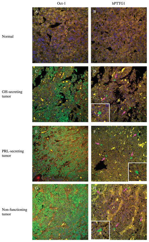
Figure 4.
Oct-1 and hPTTG1 immunoreactivity in pituitary tumor specimens. Oct-1 and hPTTG1 protein immunoreactivity were detected using fluorescence immunohistochemistry. Panel A and B show negative staining of both Oct-1 and hPTTG1 in normal tissues; compared with normal tissue, panels C–H show high Oct-1 and hPTTG1 expression in GH-secreting, PRL-secreting, and non-functioning pituitary tumors respectively. Pink arrows indicate positive hPTTG1 staining cells. Size: 375×375 μM. Green signal: staining of Oct-1 or hPTTG1; blue signal: DAPI nuclear staining; yellow signal: autofluorescence background.

