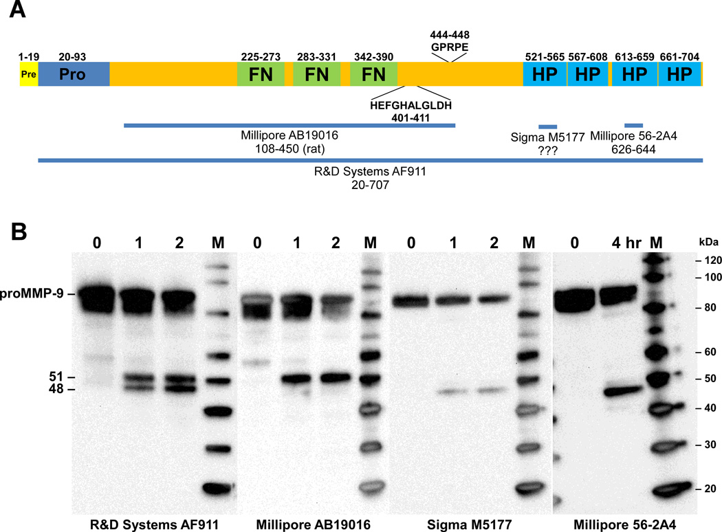
Fig. 3.
Western blot analysis of proMMP-9 cleavage products. (A) Schematic diagram of preproMMP-9 depicting the locations of the fibronectin (FN), zinc-binding catalytic (HEFGHALGLDH), and hemopexin (HP) domains. The MMP-9 immunogens used to generate the MMP-9 antibodies used for western analysis are indicated by the horizontal lines (B) Recombinant proMMP-9 was incubated with activated KLK7 for the indicated times and the reaction products were loaded in multiple lanes of 4–12% Bis-Tris gels. Following electrophoresis, the products were transferred to PVDF membranes and detected with the indicated MMP-9 antibodies. MagicMarkXP (Invitrogen) protein standards (M) were used to determine the size of the MMP-9 products. Sizes of the protein markers (kDa) are indicated on the right.

