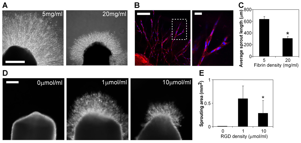
Figure 1. Decreased adhesive ligand density enhances angiogenic sprouting.
(A) Sprouting of chick aortic rings embedded in 5 mg/ml (left) versus 20 mg/ml (right) fibrin in EGM-2 medium at 48 hours, bar=500 µm. (B) Endothelial cells in sprouts labeled with lectin (red) and Hoechst 33342 (blue), with region outlined by dotted line magnified on the right. Bar (left image), 100 µm; bar (right image), 20 µm. (C) Quantification of average sprout length for chick aortic rings in 5 mg/ml versus 20 mg/ml fibrin. Graph represents means±SEM (n=4), with each experiment averaged over at least 3 aortic rings. (D) Sprouting of chick aortic rings embedded in PEGDAAm gels with 0 µmol/ml adhesive RGDS + 10 µmol/ml non-adhesive RGES (left), 1 umol/ml RGDS + 9 umol/ml RGES (middle), and 10 umol/ml RGDS + 0 umol/ml RGES (right) in EGM-2 medium at 48 hours, bar=200 µm. (E) Quantification of sprouting area for chick aortic rings in PEGDAAm gels of varying RGDS density. Graph represents means±SD (n=8). *, p<0.05, compared to 1 umol/ml RGDS, as calculated by one-way ANOVA and post-hoc Tukey’s HSD test.

