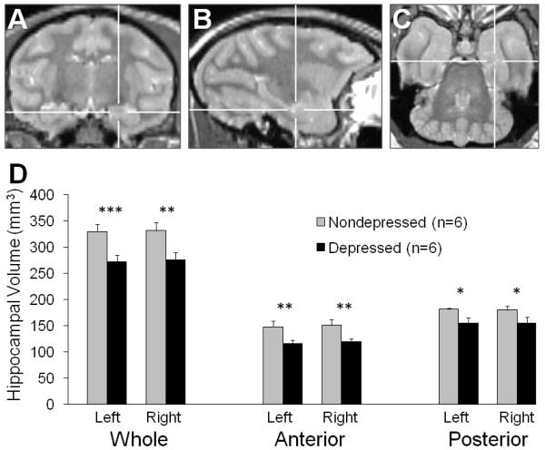
Figure 2.

Hippocampal (HC) volume in behaviorally depressed monkeys. (A – C) The anterior HC was delineated from the posterior HC by the presence of the uncus. The cross-hairs indicate the same position in all three proton-density images. (A) Coronal: The last slice in which the uncus is present marks the caudal boundary of the anterior HC. (B) Sagittal: The anterior HC appears to the right of the vertical cross-hairs. (C) The anterior – posterior boundary of the HC as viewed in the axial plane. (D) Bilateral whole, anterior and posterior HC volumes were all smaller in depressed compared to nondepressed animals. *p < 0.05; **p ≤ 0.01; ***p ≤ 0.001.
