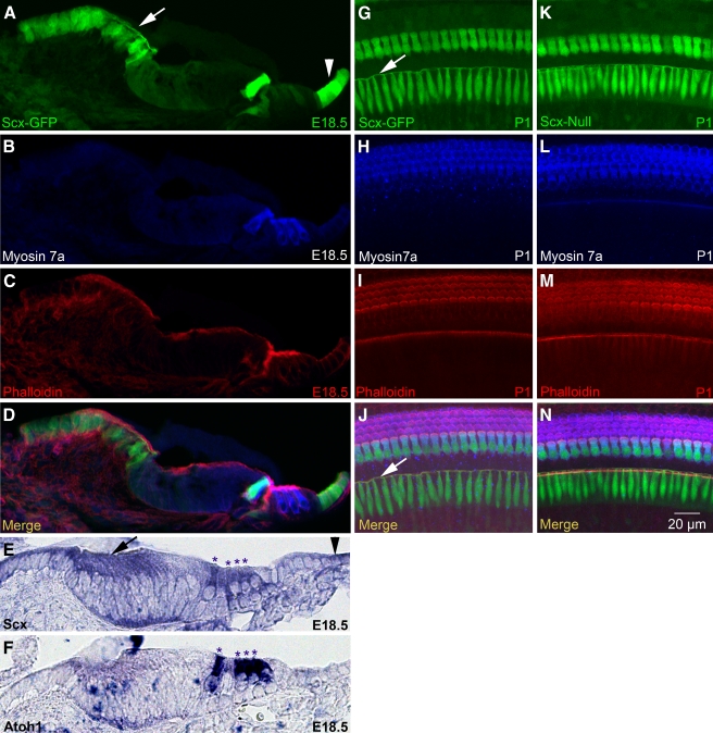
FIG. 1.
Scx is expressed in interdental cells, sensory hair cells, and Claudius cells. A–D Confocal microscopy of a cryostat section from the E18.5 Scx-GFP organ of Corti labeled with myosin 7a (B) and phalloidin (C). Panels A–C are merged in D. E, FIn situ hybridization for Scx (E) and Atoh1 (F) mRNA in cryostat sections of the E18.5 wild-type organ of Corti. Asterisks indicate apices of inner and outer hair cells. G–N Confocal microscopy of whole mount Scx-GFPG–J and Scx-nullK–N organs of Corti labeled with myosin 7a H, L and phalloidin I, M. Panels G–I are merged in J, and panels K–M are merged in N. Arrows indicate interdental cells and the arrowheads indicate Claudius cells. The cryostat section in A–D is from a more basal location in the cochlea than the sections in E and F. Scale bar in N applies to all panels.

