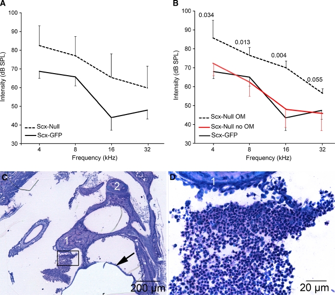
FIG. 2.
Middle ear infection correlates with elevated hearing thresholds in affected Scx-null mice. A The average auditory brainstem response thresholds in sound pressure level (dB SPL) as means ± standard deviation (SD) are plotted for Scx-null (n = 26 ears; 13 mice) and Scx-GFP (n = 20 ears; 10 mice) mice at 4, 8, 16, and 32 kHz. Auditory thresholds in Scx-null mice are statistically significantly elevated at all four frequencies tested (t > 2.02; p < 0.001). B The average auditory brainstem response thresholds (dB SPL ± SD) are plotted for Scx-null mice with otitis media (OM; dashed line, n = 4 ears from four mice) versus Scx-null mice without otitis media (red line; n = 5 ears from five mice) at 4, 8, 16, and 32 kHz. The average ABR thresholds of Scx-null mice with otitis media are statistically significantly elevated at three of the four frequencies tested (t test, p values indicated on the graph) compared to Scx-null mice without otitis media. The ABR thresholds of Scx-null mice without otitis media (B; red line) are not statistically significantly different from Scx-GFP mice (B, black line; data from A) at all frequencies tested (t > 2.07, p > 0.12). C, D Methylene blue/basic fuchsin-stained plastic thin sections of a representative Scx-null middle ear that presented with white, viscous fluid behind the tympanic membrane upon postmortem analysis. The boxed area in C is shown at higher magnification in (D). The methylene blue-stained cells between the malleus (1) and tympanic membrane (arrow) have small nuclei with scant cytoplasm consistent with infiltrating lymphocytes. Weakly methylene blue-stained acellular debris is associated with the incus (2).

