FIG. 4.
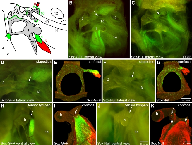
Scx is required for middle ear tendon differentiation. A The mammalian middle ear consists of three delicate bones and associated ligaments and tendons. The manubrium (m) of the malleus (1) is inserted into the tympanic membrane or ear drum (see also Fig. 2C, arrow). The incus (2) articulates with the malleus and the stapes (3) at the malleoincudal (6) and incudostapedial (7) joints, respectively, where ligaments provide structural integrity. The footplate of the stapes rests on the oval window of the cochlea where it is stabilized by the annular ligament (10). The tensor tympani muscle (5) inserts into a tubercle (11) adjacent to the head (h) of the malleus through the tensor tympani tendon (4). The stapedius muscle (9) inserts into a tubercle on the posterior arch of the stapes through the stapedial tendon (8). The round window of the cochlea (12), the stapedial artery (13), and the bony labyrinth at the level of the middle turn of the cochlea (14) are prominent anatomical landmarks in this lateral view. All middle ear tendons and ligaments express Scx and green shading schematically summarizes Scx-GFP expression. A patch of Scx-GFP-expressing tissue is localized to the arms of the malleus where they associate with the dorsal wall of the middle ear cavity. A small focus of Scx-GFP-expressing tissue is also present between an arm of the incus and the posterior wall of the middle ear cavity. P posterior; V ventral. These numerical and alphabetic labels are used in subsequent panels (B, C) Lateral view of the Scx-GFP (B) and Scx-null (C) middle ears imaged in combination bright field/epifluorescence (BF/EPI) mode (see Materials and methods). The stapedial tendon insertion point (arrow) on the tubercle of the posterior arch of the stapes is indicated. The tensor tympani tendon located behind the malleus (1) robustly expresses Scx-GFP in wild type (B) but not the null (C) middle ear. Scx-GFP is not detectable in the middle ear mucosa. Scx-GFP expression in the cochlea is detectable through the bony labyrinth in the wild type (14) and null inner ears. Scale bar in (C) applies to (B). (D, E) The stapedial tendon insertion into the tubercle on the posterior arch of the stapes in the Scx-GFP middle ear imaged by BF/EPI (D) and laser confocal (E) microscopy. The tenocytes in the stapes tendon robustly express GFP that is detectable in the whole mount image as a feathered array of green fluorescence terminating at the posterior arch of the stapes (D, arrow). The excised stapes was dissected from its associations with the oval window and incus prior to immunohistochemical detection of Myo32 and confocal analysis. The stapes tendon insertion into the tubercle and its association with a fragment of the Myo32+ stapedius muscle is shown in (E). The posterior arch of the stapes in (E) was fractured during excision or tissue processing (F, G) Scx-GFP expression is weakly detectable in the BF/EPI (F) and laser confocal (G) images of the stapes of Scx-null mice. The stapedius is inserted into the stapes by a GFP+ tendon (F, arrow and G; see also C, arrow). The stapedial tendon in Scx-null mice is dramatically shorter than the tendon in the wild type Scx-GFP middle ear (compare GFP+ tendon in E and G). The foreshortened stapedius tendon is associated with a fragment of the Myo32+ stapedius muscle in the Scx-null middle ear (G). Scale bar in G applies to (E). (H, I) The tensor tympani tendon insertion into the tubercle of the malleus in the Scx-GFP middle ear imaged by BF/EPI (H) and laser confocal (I) microscopy. The tenocytes in the tensor tympani robustly express Scx-GFP (H), and it is this intense expression that is detectable behind the malleus in the lateral view of the Scx-GFP middle ear in panel B. The matrix that anchors the tendon to the tubercle is GFP negative (H, arrow). The excised malleus was dissected from its associations with the ear drum and incus, and the bony arms were trimmed prior to immunohistochemical detection of Myo32 and confocal analysis. Myo32 expression in the tensor tympani (I; red) demonstrates the extensive association of the GFP+ tendon with the muscle and the absence of GFP expression proximal to the insertion point on the tubercle (I, arrow). (J, K) Scx-GFP expression is weakly detectable in BF/EPI (F) and laser confocal (G) images of the malleus in Scx-null mice. Confocal analysis detects a small complement of diffusely arrayed, weakly GFP+ tenocytes (K, arrowheads) associated with the Myo32+ tensor tympani. The tensor tympani inserts into the malleus by a foreshortened, stubby cord of matrix that is GFP negative (J, arrow and Fig. 6). The reduced expression of Scx-GFP in the Scx-null tensor tympani tendon accounts for the inability to detect GFP expression behind the malleus in the BF/EPI image (J) and in the lateral view of the Scx-null middle ear (C). Scale bar in (J) applies to (D, F, H). Scale bar in (K) applies to (I).

