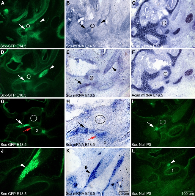
FIG. 5.
Scx-GFP fluorescence accurately reports Scx mRNA expression in middle ear tendons and the incudostapedial ligament. White and black arrows indicate the developing stapedius tendon, and arrowheads indicate the tensor tympani tendon in all panels. A, D, G, J, I, LScx-GFP expression at E14.5 (A), E16.5 (D), E18.5 (G, J), and P0 (I, L) in cryostat sections of the Scx-GFP middle ear. C, FAcan expression in chondrocytes of the nascent stapes at E14.5 (C) and E16.5 (F) and chondrocytes in the developing cartilaginous capsule of the inner ear. The circle outlines the obturator foramen of the stapes through which the stapedial artery passes. Acan provides an anatomical framework to interpret Scx-GFP and mRNA expression in (A, B, D, E). H, KScx-mRNA expression in the stapedius tendon (H, black arrow) and tensor tympani tendon (K, arrowhead) at E18.5. The narrow band of Scx-GFP expression (G, red arrow; 2, incus) in the incudostapedial ligament correlates with Scx mRNA expression (H, red arrow). Scale bar in (K) applies to (H, J); scale bar in (L) applies to all other panels.

