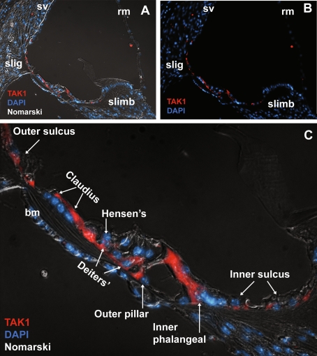
FIG. 6.
Down-regulation of TAK1 in the P16 cochlea. A, B By P16, TAK1 immunostaining was down-regulated in the stria vascularis, Reissner’s membrane, spiral limbus and both inner and outer hair cells. TAK1 labeling was present in the fibrocytes in the spiral ligament (sl) and supporting cells of the organ of Corti. C Higher magnification reveals that TAK1 immunostaining was limited to the supporting cells of the organ of Corti as well as the Claudius cells. TAK1 immunolabeling was also seen in the cells of the inner and outer sulcus. Cell nuclei are counterstained with DAPI (blue). The gamma was adjusted in these images in order to reduce nonspecific background autofluorescence.

