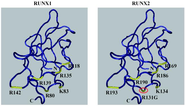
Figure 5. Three-dimensional structure of RUNX1 and RUNX2 runt domain.
The three-dimensional structures of runt domains from RUNX1 and RUNX2 were annotated based on amino acid numbering for each protein. Red circle indicates the novel missense mutation site (R131G) and yellow color points to DNA binding residues. The three-dimensional structures were obtained from NCBI (CDD pfam00853).

