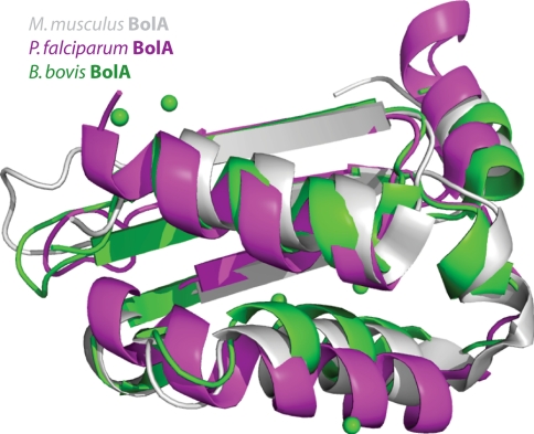
Fig. 4.
Overlay of NMR solution structures of BolA-like proteins from M. musculus (gray, PDB ID 1V9J [42]), P. falciparum (magenta, Buchko, G.W. et al. unpublished) and the crystal structure of a BolA-like protein from B. bovis solved by iodide ion SAD (green, PDB ID 3O2E). Iodide ions are shown as green spheres. For simplicity only the ordered regions are shown

