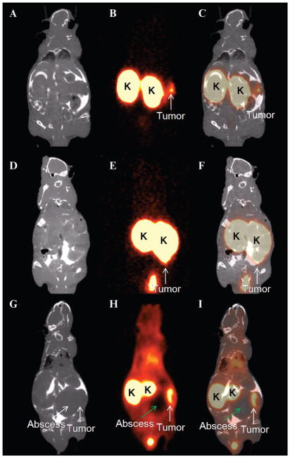
Figure 6.
Coronal slices of PET images (B, E, H) of three mice with intraperitoneal tumors after injection with 68Ga-DOTA-MG0. PET images were acquired 1 hour postinjection. CT images (A, D, G) were acquired (after intraperitoneal injection of ioiversol [Optiray 300]) and fused with the PET images (C, F, I) for anatomic correlation. Tumors are clearly visible (white arrow), and the kidneys (K) exhibit high uptake of 68Ga-DOTA-MG0. The abscess (green arrow) present in the abdomen of one mouse (G–I) showed no uptake of 68Ga-labeled MG0. Images were acquired 28 to 35 days after tumor inoculation.

