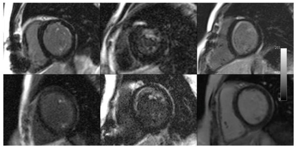
Figure 1.
Late gadolinium enhancement (LGE) images from all 6 patients. The bottom right image shows normal myocardium. The left and middle columns show enhancement in both papillary muscles, with enhancement in the anterior papillary muscle. There are varying degrees of anterior sub-endocardial LGE in the 5 abnormal images.

