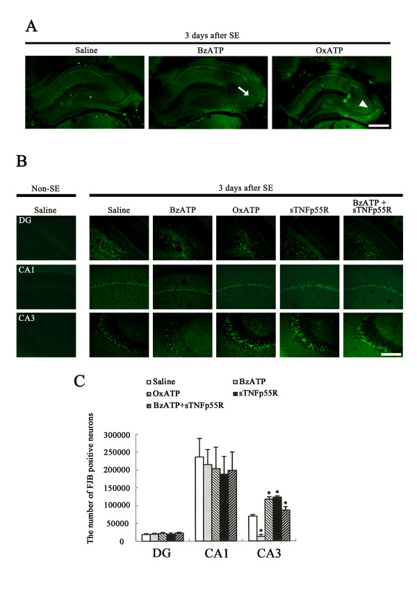
Figure 5.
Effect of BzATP, OxATP, sTNFp55R, and BzATP+sTNFp55R infusions on SE-induced neuronal death. (A) Representative photographs of FJB staining following SE. As compared to saline-infusion, BzATP infusion attenuates neuronal damage in the CA3 region (arrows), while OxATP infusion worsens it (arrowheads). (B) SE-induced neuronal damages in dentate gyrus, and in the CA1 and CA3 regions 3 days after SE. Bar = 100 μm. (C) Quantitative analysis of neuronal damage in dentate gyrus, and in the CA1 and CA3 regions 3 days after SE (mean ± S.E.M). BzATP infusion alleviates SE-induced neuronal damage only in the CA3 region. However, the other treatments increase SE-induced neuronal damage. *Value is significantly different from saline-infused animals, p < 0.05.

