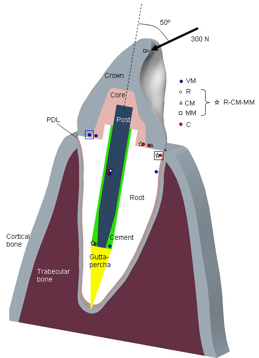
Figure 1.

Section of the tooth model with position of failure initiation points for the different criteria. Longitudinal section of the geometrical model simulating a typical endodontic restoration of a maxillary central incisor including the elements: bone (cortical and trabecular components), periodontal ligament (PDL), root, gutta-percha, post, cement, core and crown. Position and orientation of the load applied for the FE model to simulate a real biting force. Marks indicate position of the points with the lowest SF in each component for the different criteria considered. A common mark was used for R, CM and MM criteria when they predicted the critical point in the same position. Big squares indicate the area predicted for failure initiation by the different criteria (VM: blue square, R-CM-MM-C: black square).
