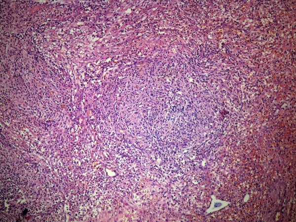
Figure 2.
Macrophages are forming aggregates, while the rest of the inflammatory cells are interspersed in a stroma with abundant fibroblasts and collagen bundles. The inflammation partially extends to the adjacent liver parenchyma. The aggregate of macrophages is located in the lower right part of the image (hematoxylin and eosin, × 400).

