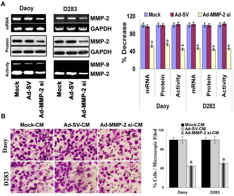
Figure 2. Ad-MMP-2 si-infection inhibits MMP-2 expression in medulloblastoma cells and decreases human umbilical cord blood stem cell (hUCBSCs) migration towards tumor cells.

Medulloblastoma cells (Daoy and D283 cells) were treated with mock (PBS Control), 50 MOI of Ad-SV or Ad-MMP-2 si for 24 h, and conditioned medium and cells were collected. (A) Top panel: Total RNA was extracted and semi-quantitative RT-PCR was performed for MMP-2; GAPDH was served as a control for RNA quality. Middle panel: MMP-2 levels were determined in the total cell lysates by western blot analysis using anti-MMP-2 antibody. GAPDH served as a loading control. Bottom panel: MMP-2 gelatinolytic activity was determined in the conditioned medium by gelatin zymography. Band intensities were quantified by densitometric analysis using ImageJ software (National Institutes of Health). The levels of MMP-2 mRNA, protein and gelatinolytic activity were normalized to levels in mock-transfected cells. Columns: mean of triplicate experiments; bars: SD; *p<0.05, significant difference from mock transfected control cells. (B) hUCBSCs migration towards tumor cell conditioned medium from medulloblastoma cells infected with Ad-MMP-2 si was studied in a transwell migration assay. Conditioned medium collected from mock, Ad-SV and Ad-MMP-2 si-infected tumor cells were designated as mock-CM, Ad-SV-CM and Ad-MMP-2 si-CM, respectively. The cells that had migrated to the lower side of the membrane were photographed under a light microscope at 20× magnification. Percentages of migrated cells were quantified by counting five fields in each condition. Results are representative of three independent experiments. Columns, mean of three experiments; bars, SD.
