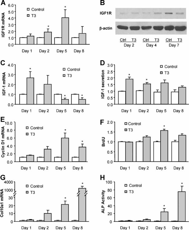
Fig. 1.

Thyroid hormone treatment increases Igf1r expression in rat growth plate chondrocytes. (A, C) Quantitative real-time RT-PCR analysis of Igf1r and Igf1 mRNA expression in pellet cultures of growth plate chondrocytes treated with 100 ng/mL of T3 for 1 to 8 days. The expression in T3-treated cells was normalized to the expression in control cells. (B) Immunoblotting of IGF1R protein from whole-cell lysates of growth plate chondrocytes treated with or without T3 (100 ng/mL) for 2 to 7 days. Actin was used as an internal control. (D) Detection of IGF-1 protein in culture medium of growth plate chondrocytes treated with or without T3 for 1 to 8 days. (E, F) Cyclin D1 mRNA and BrdU incorporation in growth plate cells treated with or without T3 for 1 to 8 days. (G, H) Col10a1 mRNA and alkaline phosphatase activity in growth plate chondrocytes treated with or without T3 for 1 to 8 days. *p < .05 versus control cells.
