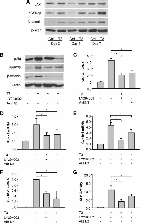
Fig. 5.

Thyroid hormone promotes IGF-1-activated PI3K/Akt signaling and PI3K/Akt-dependent β-catenin signaling in growth plate chondrocytes. (A) Immunoblotting of phosphorylated Akt (pAkt), phosphorylated GSK-3β (pGSK-3β), and active β-catenin protein from whole-cell lysates of growth plate chondrocytes treated with or without T3 (100 ng/mL) for 2 to 7 days. Actin was used as an internal control. (B) Immunoblotting of pAkt, pGSK-3β, and β-catenin protein of growth plate chondrocytes treated with T3 for 5 days in the presence or absence of PI3K signaling inhibitor LY294002 (20 µM) and Akt signaling inhibitor Akti1/2 (1 µM). (C–E) Wnt4 mRNA (C), Runx2 mRNA (D), and cyclin D1 mRNA (E) expression of growth plate chondrocytes treated with T3 for 5 days in the presence or absence of LY294002 and Akti1/2. *p < .05 versus the cells treated with T3 alone. (F, G) Col10a1 mRNA (F) and ALP activity (G) of growth plate cells treated with T3 for 5 days in the presence or absence of LY294002 and Akti1/2. *p < .05 versus the cells treated with T3 alone.
